10+ block diagram ecg
Critical Decisions in Emergency and Acute Care Electrocardiography 1e 2009. The recording of a heart beat an ECG may be corrupted by noise from the AC mainsThe exact frequency of the power and its harmonics may vary from moment to moment.

Adas1000 Datasheet And Product Info Analog Devices
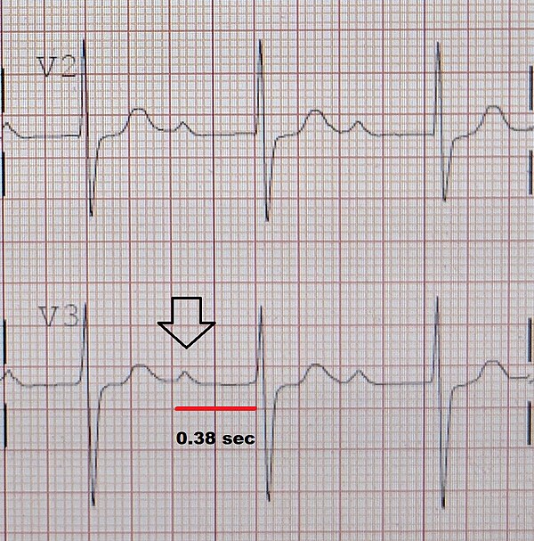
So the height and depth of these signals are a measurement for the voltage.

. Some calipers can be as simple as a compass with inward or outward-facing points but no scale. Single-chamber pacemakers the most commonly used devices deliver electrical impulses to the right atrium upper chamber of the heartThe sinus node a cluster of cells in the right atrium is the hearts natural pacemaker. PJC Premature junctional complex B.
Time Series Classification TSC is an important and challenging problem in data mining. If this is not set at 10 mm there is something. Am Heart J 19305685.
Mattu A Brady W. In electrocardiography left axis deviation LAD is a condition wherein the mean electrical axis of ventricular contraction of the heart lies in a frontal plane direction between 30 and 90. When you are taking anatomy and physiology you will be required to identify major muscles in the human body.
This is reflected by a QRS complex positive in lead I and negative in leads aVF and II. ECGs for the Emergency Physician Part I 1e. The block diagram in Fig.
ECG changes and extension of the infarction depend heavily on the site of the occlusion. Get all these features for 6577 FREE. Atrial fibrillation AF or A-fib is an abnormal heart rhythm arrhythmia characterized by rapid and irregular beating of the atrial chambers of the heart.
The cardiac cycle is the performance of the human heart from the beginning of one heartbeat to the beginning of the next. Adult and Pediatric 6e 2008. Third-degree atrioventricular block AV block is a medical condition in which the nerve impulse generated in the sinoatrial node SA node in the atrium of the heart can not propagate to the ventricles.
What type of arrhythmia is labeled by the arrows. Essay Help for Your Convenience. The more proximal the occlusion the greater the infarction and the more pronounced ECG changes.
It may also start as other forms of arrhythmia such as atrial flutter that then transform into AF. The ECG Made Practical 7e 2019. ECG Pocket Brain Expanded 6e 2014.
One during which the heart muscle relaxes and refills with blood called diastole following a period of robust contraction and pumping of blood called systoleAfter emptying the heart immediately relaxes and expands to receive. The tips of the caliper are. In the diagram opposite - a glass tube b muscle c sliver wire d brass wire e drop of water f investigators hand.
The ECG EES includes two epidermal electrodes an instrumentation amplifier analog filters an inverting amplifier. This quiz requires labeling so it will test your knowledge on how to identify these muscles latissimus dorsi trapezius deltoid biceps brachii triceps brachii brachioradialis pectoralis major serratus anterior rectus. Brady WJ Truwit JD.
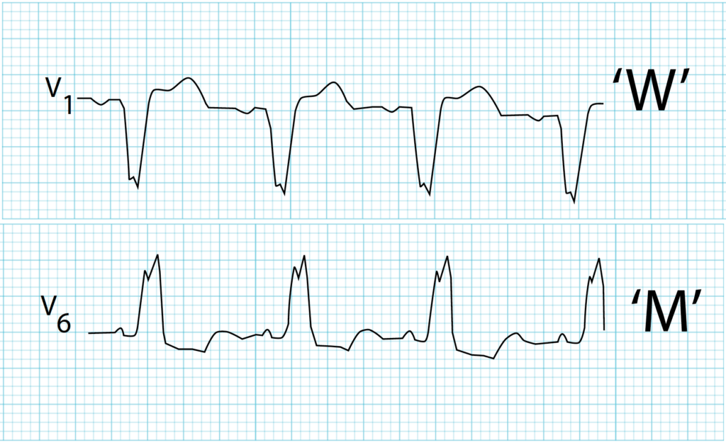
Left Bundle Branch Block. Smiths ECG Blog for illustration on how to. Dual-chamber pacemakers are used when the timing of the chamber contractions is misalignedThe device corrects this by delivering.
Smiths ECG Blog I have found the Mirror Test to be an extremely helpful visual aid for recognizing the special shape of anterior ST depression seen in association with acute posterior OMI See My Comment in the January 3 2022 post in Dr. It consists of two periods. Because the impulse is blocked an accessory pacemaker in the lower chambers will typically activate the ventricles.
One way to remove the noise is to filter the signal with a notch filter at the mains frequency and its vicinity but this could excessively degrade the quality of the ECG since the. With the increase of time series data availability hundreds of TSC algorithms have been proposed. To discuss your healthcare needs call us on.
It is a unique ECG phenomenon consisting of complexes formed by the blurring together of QRS and T-wave as a result of extreme ST-Deviation. This ladder diagram illustrates which arrhythmia. Some of the causes include normal variation thickened left.
1G summarizes the system architecture and overall wireless operation of our systems. Electrocardiography is the process of producing an electrocardiogram ECG or EKG a recording of the hearts electrical activity. 1st degree AV block.
We cover any subject you have. A caliper British spelling also calliper or in plurale tantum sense a pair of calipers is a device used to measure the dimensions of an object. These electrodes detect the small electrical changes that are a consequence of cardiac muscle depolarization.
At the beginning of every lead is a vertical block that shows with what amplitude a 1 mV signal is drawn. The hearts electrical activity begins in the sinoatrial node the hearts natural pacemaker which is situated on the upper right atriumThe impulse travels next through the left and right atria and summates at the atrioventricular nodeFrom the AV node the electrical impulse travels down the bundle of His and divides into the right and left bundle branches. As Ive emphasized on many occasions in Dr.
These complexes manifest in contiguous ECG leads corresponding with coronary anatomy and represent transmural ischemia. Choose from the following responses to interpret this ECG. The Institute comprises 33 Full and 13 Associate Members with 12 Affiliate Members from departments within the University of Cape Town and 12 Adjunct Members based nationally or internationally.
Chous Electrocardiography in Clinical Practice. Many types of calipers permit reading out a measurement on a ruled scale a dial or a digital display. Among these methods only a few have considered Deep Neural Networks DNNs to perform this task.
1091 The best writer. There are several potential causes of LAD. It is an electrogram of the heart which is a graph of voltage versus time of the electrical activity of the heart using electrodes placed on the skin.
Muscle anatomy quiz for anatomy and physiology. ST-segment elevations may be present in leads V1V6 and frequently aVL I the latter two may be affected because the diagonals given off by the LAD supplies. 9 Color coding of the ECG leads.
Surawicz B Knilans T. Personal 0808 271 8573 Members. Receive your papers on time.
Bundle branch block with short P-R interval in healthy young people prone to paroxysmal tachycardia. It often begins as short periods of abnormal beating which become longer or continuous over time. 12-lead ECG library A brief history of electrocardiography from 1600 onwards.
Set the deadline and keep calm. 常識を超えるThe ICE 27 冷感寝具は もう必要ありません 夏の快眠温度で感動の寝落ち 快適な温度2733を長く持続する夏の寝具The ICE 27ザアイス27. This is surprising as deep learning has seen very successful applications in.
Any Deadline - Any Subject. Shark Fin is an electrocardiographic sign of acute coronary occlusion.

Automated Ecg Interpretation Wikiwand
What Effect Does Holding Breath Have On An Ecg Signal Quora

What Does St Segment Signify In An Ecg Quora
What Are The Qrs Morphology In Ecg Signals Quora
What Are The Characteristics Of An Atrial Fibrillation Ecg Quora

Adas1000 Datasheet And Product Info Analog Devices

Adas1000 2 Datasheet And Product Info Analog Devices
Our Team Ecg Weekly
Carolinas Ekg Blog Cmc Compendium

Sinus Rhythm Wikiwand
Carolinas Ekg Blog Cmc Compendium

First Degree Atrioventricular Block Wikiwand

Left Bundle Branch Block Wikiwand

Adas1000 Datasheet And Product Info Analog Devices

Ecg Interpretation Ecg Blog 266 74 Dewinter T Waves Or Post Mi
Carolinas Ekg Blog Cmc Compendium

Adas1000 1 Datasheet And Product Info Analog Devices